Orofacial pain conditions - Pain and oral mucosa
Pain of the oral mucosa is a common accompanying symptom of various oral mucosal lesions caused by local and systemic diseases. Pain of the oral mucosa is usually associated with a known cause of tissue damage, e.g. mucosal ulcer or erosion, and it generally responds to adequate treatment and dissolves after healing. Chronic pain, on the other hand, persists months and years after apparent tissue healing, and attempts to alleviate pain are challenging. Neuropathic pain occurs due to damage neurogenic structures in the peripheral and/or the central nervous system. It may occur in the absence of any obvious noxious stimuli, and in the oral mucosal, the pain is often described as tingling and burning. In the oral cavity, burning mouth syndrome (BMS) is presently considered to have neuropathic background. It is important for dental practitioners to have a clear understanding of the various diseases that can cause oral mucosal pain to provide appropriate care to patients. This paper focuses on the most common local and systemic diseases that can cause oral mucosal pain with respect to their clinical features and management.
Patients with intermittent or persistent, painful sensations in the oral mucosa often represent a substantial clinical challenge with regard to diagnosis and management. The oral cavity is one of the most densely innervated parts of the body, and it also has an extensive sensorimotor representation in the central nervous system (CNS). The rich somatosensory supply, in terms of peripheral receptors, is related to the important role of the mouth in oral sensorimotor control in eating, drinking, swallowing and speaking, and also in the large variety of oral sensations, including pain (1 - 3).
Acute oral mucosal pain especially occurs in association with inflammation, oral surgery or accidental injury. Most conditions with acute pain can be treated, and usually subside when healing of the tissue has taken place. Chronic pain, on the other hand, persists months and years after apparent tissue healing, and attempts to alleviate pain often fail (4). Moreover, chronic pain conditions also appear to be associated with structural and functional alterations in the CNS (5). Accordingly, early and appropriate diagnosis and management of acute pain is important in order to avoid that acute pain turn into a chronic pain condition with impaired quality of life and risk of psychological morbidity like anxiety and depression (4).
Oral mucosal pain is often characterised by a burning, stinging or sore sensation. Various mucosal lesions like ulcers, erosions and blisters are common causes of oral mucosal pain, and these lesions can occur due to a large variety of local mucosal and systemic diseases, of which some may be iatrogenically induced, e.g. due to surgical trauma, certain medications or radiotherapy to the head and neck region (Figures 1 and 2, Box 1). However, pain of the oral mucosa may also occur in the absence of any findings like for example neuropathic pain caused by damage of the peripheral and/or central nervous system, or be of psychogenic origin.
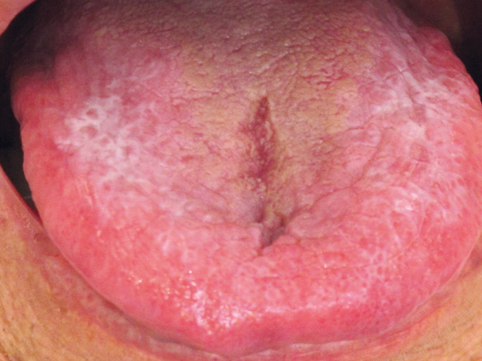
Figure 1. 74-year old female with oral lichen planus. She was referred due to itching and burning sensation of the dorsal part of her tongue, especially in relation to intake of spicy and acidic food. Note the reticular whitish striae on the tongue. She also had a history of small ulcerations on the marginal part of the tongue.
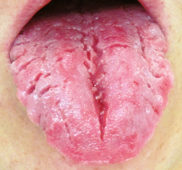
Figure 2. 32-year old female with deep fissures on the dorsal and marginal parts of her tongue as well as a geographic tongue. She complained of intermittent tingling and burning sensation on her tongue.
Box 1
A large variety of local mucosal and systemic diseases are associated with pain due to formation of ulcers or erosions. Theses lesions differ with regard to extension in the oral mucosa:
A mucosal ulcer is defined as a loss of surface tissue and disintegration and necrosis of epithelial tissue. It involves damage to both epithelium and lamina propria. It penetrates the epithelial-connective tissue border, and has its base at a deep level in the submucosa, and in some cases even within the muscle or periosteum
-
A mucosal erosion is defined as a superficial break on the mucous membrane with loss of the superficial epithelial cells and minor damage to the underlying lamina propria.
It may reach the basement membrane.
This paper presents an overview of the most common local and systemic diseases that can cause oral mucosal pain, categorised according to their clinical characteristics and management.
Oral mucosal pain mechanisms
Oral mucosal pain is often associated with tissue damage and concomitant inflammation. Pain occurs as a result of activation and/or sensitisation of nociceptors on peripheral nerve fibres by inflammatory mediators and by mechanical and thermal stimuli. Two types are distinguished based on afferent fibre morphology. A-delta fibres are myelinated and relatively fast-conducting, but slower than mechanoreceptors. They provide fast and sharp sensations of pain to noxious stimulation. C-fibres are unmyelinated and slow-conducting. They are responsible for diffuse, dull, slow aching pain (6). They are primarily located in the connective tissue and around the subepithelial capillary plexus. The activating inflammatory mediators include bradykinin, serotonin, glutamate, and H+; the sensitising mediators include prostaglandins, serotonin, noradrenaline, nitric oxide, and nerve growth factor (6). During inflammation, nociceptors display a lower threshold for stimulation-induced pain or an increased sensitivity to noxious stimuli, a condition known as hyperalgesia.
Oral mucosal pain may also occur in the absence of evident pathology or explanation, e.g. previous trauma, and is termed «idiopathic» pain. Neuropathic pain occurs as a result of damage neurogenic structures in the peripheral and/or the central nervous system (7), as there is not always a clear history of nerve injury, e.g. from local anaesthetic or surgery. After the injury, which may include direct nerve damage or tissue inflammation, the peripheral afferent nerve fibres react with increased excitability and spontaneous tonic activity. This may release permanent, neuroplastic alterations in the central neurons that contribute to maintain the nociceptive activity (8).
Chronic neuropathic pain conditions
Conditions that may be associated with chronic neuropathic pain in the oral mucosa include post traumatic trigeminal neuropathy, trigeminal post herpetic neuralgia, and burning mouth syndrome (BMS), of which the latter is the predominant one.
Burning mouth syndrome
BMS, sometimes also called stomatodynia or glossodynia, is defined as burning or painful sensations of oral mucosa with no clinical signs of pathology or identifiable medical or dental causes (9).
Considerable progress has been made in the understanding of BMS pathophysiology. An important step in this process has been the differentiation between primary BMS and what could be called secondary BMS as its symptoms mimic primary BMS. However this condition is due to clinically identifiable etiological factors. A sensation of oral burning can be associated with a large variety of systemic or local conditions of which some are reviewed in this paper (10 - 12). In these cases, treatment of the underlying cause will often alleviate the sensory symptoms. Several local and systemic factors need therefore to be taken into consideration before the diagnosis of primary (idiopathic) or secondary BMS can be made.
Reported prevalence rates of BMS in the general populations vary from 0.7 % to 4.6 % (10). The prevalence of BMS increases with age, with the highest prevalence (12 %) in women aged 60 - 69 (13). Very little is known of the prognosis of BMS but there is anecdotal evidence that BMS symptoms are long lasting.
Clinical features of BMS
Burning pain of the oral mucosa is the cardinal feature of primary BMS. The intensity of pain varies from mild to severe. It is most often experienced at more than one oral site, the anterior part of the tongue, the anterior hard palate and the lips being most frequently affected. Pain is most often bilateral and symmetrical. Most patients experience negligible symptoms on awakening, and symptoms build up over the day, being most intense in the evening, but the pain only seldom disturbs sleep. Some patients, however, experience constant symptoms throughout the day, while others only have intermittent symptoms (14,15).
More than half of the patients complain of xerostomia (11,14). Furthermore, up to 70 % of BMS patients report taste disturbances, such as alterations in taste perception and/or dysgeusia (usually bitter or metallic), or phantom tastes (14,15).
Pathophysiology of BMS
Recent studies have revealed that several neuropathic, mainly subclinical mechanisms act at different levels of the somatosensory system and contribute to the pathophysiology of primary BMS (12,16). Some studies have demonstrated that small-fibre mediated neuropathy is a common finding in BMS and that approximately a fifth of BMS patients show subclinical trigeminal nerve lesions (10,14). Furthermore, several electrogustatometric studies have reported evidence for chorda tympani hypofunction in BMS (17).
Central nervous system pathology seems also to be involved in the generation of BMS symptoms. A study on cerebral reorganization demonstrated altered grey and white matter volumes and altered functional connectivity patterns in BMS (18). Two positron emission tomography (PET) studies have demonstrated a decline in endogenous dopamine levels in BMS, suggesting deficiencies in central pain modulation (19,20).
Management of BMS
First of all, it is important that dentists recognize the syndrome, give a credible explanation of the current understanding of the pathophysiological mechanisms of BMS, and reassure the patient of its benign nature. Reassurance alone can suffice in some cases, and lead to symptom resolution and /or better coping. Some patients experience pain alleviation while eating, chewing gum, sucking pastilles, drinking cold beverages, or by avoiding spicy foods (15). As regards the actual therapies for BMS there is some evidence from RCTs for the effectiveness of topical clonazepam and antioxidant alpha-lipoid acid, as well as cognitive behavioural group therapy (21). The topical use of clonazepam (1 mg three times a day) improves the symptoms in about two thirds of the patients (presumably in patients whose symptoms is due to peripheral neuropathy), and is the first choice for treatment when medication is needed. As the present understanding of BMS pathophysiology suggest neuropathic involvement, also drugs that are effective in other neuropathic pain conditions, such as tricyclic antidepressants or gabapentinoids, can be used for BMS.
Conditions causing pain due to mucosal tissue injury and inflammation
Painful oral mucosal ulcers, erosions and blisters may occur due to a large variety of diseases. Table 1 gives an overview of local and systemic diseases causing mucosal ulcers, erosions or blisters due to various inflammatory reactions, autoimmune-mediated epithelial damage (e.g. mucous membrane pemphigoid), immune deficiency, and mucosal trauma. The table also indicates principles of management.
Condition/disease |
Clinical features |
Management |
|---|---|---|
Mucocutaneous diseases |
||
Lichen planus (22,23)(see Fig. 1) |
Oral lichen planus (OLP) lesions often present bilaterally and are usually seen on the buccal mucosa, the tongue and gingiva.There are 6 types: Reticular, erosive/ulcerative, papular, plaque-like and bullous OLP. They may be present simultaneously.The reticular type is the most common one and usually asymptomatic, whereas the erosive/ulcerative types often accompanied by a burning, stinging pain. |
Topical or systemic corticosteroidsCalcineurin inhibitorsWeak evidence regarding pain relief:Topical tacromilus and pimecrolimusTopical ciclosporinTopical aloe vera |
Pemphigus vulgaris (24) |
An autoimmune disorder with deposition of mainly IgG class antibodies intercellularly as well as damage to desmosomes by antibodies directed against the extracellular domains of cadherin-type epithelial cell adhesion molecules, particularly desmoglein 3, resulting in multiple ulcers and erosions preceded by bullae, mainly on the soft palate and buccal mucosa. |
Systemic treatment including corticosteroids, immunoglobulins, rituximab, mycophenolate mofetil,methotrexate, azathioprine, and cyclophosphamide |
Mucous membrane pemphigoid (25) |
An acute or chronic autoimmune disease, occurring due to reaction to the epithelial basement membrane, causing desquamation and ulceration of the oral mucosa. The most common sites of oral involvement are the gingiva (94 %), palate (32 %), buccal mucosa (29 %), floor of the mouth (5 %), and tongue (5 %). |
Topical steroids such as clobetasolSystemic prednisoneOther immunosuppressive agents e.g. methotrexate, azathioprine, mycophenolate mofetil may be indicated. Antibiotics, e.g. tetracycline or erythromycin to control secondary infections |
Erythema multiforme (26) |
Acute onset; extensive, irregular and extremely painful areas of ulcerations with yellow base and erythematous borders on buccal mucosa, palate, dorsal and ventral surfaces of the tongue.The lips are often involved showing extensive irregular ulcerations, cracking and fissuring with blood encrustation.Erythema multiforme is usually triggered by herpes simplex infections, and occasionally by drug intake. |
Identification of triggering agent.If HSV infection, antiviral therapyIf it is an adverse drug reaction, the drug is immediately stopped. Palliative treatment: analgesics, viscous lidocaine rinses, soothening mouth rinses, soft diet, avoidance of acidic and spicy food, systemic or topical antibiotics to prevent secondary infection; systemic or topical corticosteroids in severe cases |
Lupus erythematosus(25) |
Erythematous and ulcerative lesions with white striae radiating from the center, on buccal and labial mucosa, gingiva and vermillion.The lesions can also be white or red patches or bullous. |
Systemic or topical corticosteroidsSystemic immunosuppressive agents |
Iatrogenic conditions |
||
Oral Graft versus hostDisease (27) |
Characterised by lichenoid, papular and erythematous lesions, and occasionally ulcerations and desquamation on the buccal and labial mucosa, the palate and dorsal part of the tongue. The oral lesions are often accompanied by fever, malaise, nausea, and xerostomia. The oral findings may be caused by a combination of radiotherapy, chemotherapy, immunosuppressive medications, and secondary infections. |
Systemic immunosuppressive agents |
Oral mucositis (28) |
A condition that may occur in patients who receive high-dose chemotherapy, and/or radiotherapy to head and neck cancer involving the oral cavity. Oral mucositis refers to erythematous and ulcerative lesions of the oral mucosa. The lesions are often very painful and compromise nutrition and oral hygiene as well as increase risk for local and systemic infection.The condition may also be accompanied by taste disturbances and xerostomia.Acute mucositis may progress into chronic mucositis. |
Palliative treatment:Maintenance of sufficient oral hygieneSip plenty of waterAnalgesics, viscous lidocaine rinsesLubricating, soothening mouth rinses/gelsSoft diet, avoidance of alcohol, acidic and spicy foodSystemic or topical antibiotics (antibacterial, antiviral and antifungal treatment) |
Medication-inducedreactions (26) |
Often extensive irregular ulcerations of variable depth, most commonly seen on the buccal mucosa and gingiva.Erosions as well as pemphigus-like and lichenoid lesions may also be present. |
Discontinuing use of the offending medication, which could be NSAIDs, penicillamine, pyrazolone; antihypertensives like captopril and beta-blockers; antibiotics e.g. penicillin, rifampin and cephalexin; barbiturates and hormones.Topical steroids to enhance healing |
Oral allergy syndromeand contact allergy (29) |
Oral allergy syndrome (OAS) usually occurs in patients who are allergic to pollen from trees, grasses or weeds. Fresh fruit, raw vegetables and raw nuts are common causes of OAS. The symptoms including itching sensation and/or swelling of all or part of the lips, tongue, mouth or throat, but this can on occasions be severe and also include nausea and vomiting.Dental materials, oral hygiene products and food additives may cause contact allergic reactions in the mouth with varied clinical presentation including stomatitis, lichenoid lesions, erosions, blisters and ulcerations. |
Avoidance of the allergens.Identification of cause of the allergy by patch testing.Systemic or topical corticosteroidsAntihistamine |
Local mucosal conditions |
||
Reccurent aphthous stomatitis (RAS) (30) |
RAS is characterised by recurrent bouts of solitary or multiple shallow painful ulcers with erythematous borders, at intervals of few months to few days. Ulcers are most commonly seen in the non-keratinised mucosal surfaces like labial mucosa, buccal mucosa, and floor of the mouth.Ulcers may be 2 - 5 mm in diameter (minor RAS), > 10 mm (major RAS), or herpetiform (ulcers of 2 - 4 mm arranged in crops) |
Potential systemic association with RAS must be ruled out, especially in cases with sudden development of RAS in adulthoodChlorhexidine mouth rinseTopical steroids and systemic prednisone in severe casesTetracycline oral suspension |
Geographic tongue (31)(see Fig. 2) |
Circular erythematous areas, often sharply defined by elevated, whitish border zones, located at the lateral, dorsal, anterior, and/or ventral parts of the tongue The erythematous appearance occurs due to atrophy and loss of filiform papillae lesionsAbout 30 % have oral discomfort, burning and stinging sensation on the tongue |
Symptomatic treatment:Benzydamine hydrochlorideSoothening mouth rinseTopical steroids in severe cases |
Traumatic ulcers |
Anywhere on the oral mucosaLocalised ulcers with red borders produced by accenditial biting of oral mucosa, pentration by a foreign object or irrition by a denture dental restoration orthodontic applienes |
Removal of causeHeals in 7 - 10 days unless secondarily infected |
Inflammatory bowel diseases |
||
Crohn's disease (32) |
Multifocal, linear, nodular, or diffuse mucosal thickenings seen in thelabial and buccal mucosa, and the mucobuccal folds. They may beassociated with (persistent) ulcerations.Aphtous-like ulcerations and atrophic glossitis |
Systemic treatment with immunosuppressants.Some oral ulcerating lesions may require topical corticosteroid therapy or intralesional corticosteroid injections. |
Ulcerative colitis (32) |
Scattered, clumped or linearly oriented pustules on an erythematous mucosa at multiple oral sites.Some patients exhibit oral aphthous-like lesions in addition to the pustular lesions. |
The oral lesions usually respond to the systemic treatment.Topical or systemic corticosteroids and dapsone have been used for recalcitrant oral lesions with variable effectiveness. |
Coeliac disease (33) |
Aphthous-like ulcers are commonMalabsorption of iron and vitamin B may lead to burning, stinging sensations in the tongue. |
Gluten-free dietAphthous-like lesions usually disappear or improve in patients who adhere to a gluten-free diet |
Mineral and vitamin deficienies | ||
Iron deficiency Vitamin B12 and folate deficiencies (32) |
Atrophic glossitis in which the filiform papilla of the dorsum of the tongue undergo atrophy, leaving a smooth, erythematous tongue. Other parts of the oral mucosa may also appear atrophic and red.Aphthous-like ulcers are common in severe cases.Burning, stinging sensation may precede clinically detectable oral lesionsSevere cases of vitamin B12 may also be associated with paresthesia.Predisposition to develop angular cheilitis. |
Iron and vitamin B12 and folate deficiencies often occur due to malabsorption in gastrointestinal diseases or due to pernicious anaemia.The oral manifestations are often responsive to appropriate replacement therapy. In severe cases of vitamin B12, paresthesia may persist. |
Oral infections
A number of bacterial, fungal and viral infections may also cause oral discomfort and pain due to formation of vesicles, blisters, erosions and ulcers. The most common infections and their management are described in Table 2.
The oral cavity plays a key role in the life strategy of several viruses. Mucous membranes (being wet and surfaced by live cells) are generally easier to penetrate than intact skin, and the location has obvious qualities in terms of access and transmission (34). A majority of people harbour at least one of the two herpes simplex viruses (HSV), of which type 1 is the one more often associated with the mouth (Table 2). A recent case report suggests that HSV-1 can be responsible for BMS-like symptoms (35). The patient had a high titre of the virus in saliva, and the pain disappeared upon antiviral treatment.
Bacterial and fungal infections |
Clinical features |
Management |
|---|---|---|
Acute necrotising ulcerative gingivitis/stomatitis |
Painful, bleeding gingiva characterised by necrosis and ulceration of gingival papillae and margins.Foetor ex ore |
Oral hygiene instructionsChlorhexidine mouth rinseAnalgeticsTreatment of underlying disease |
Oral candidiasis (36) |
Common fungal infection, predominantly caused by Candida albicans.Usually occurs when the oral homeostasis is disturbed; namely in relation to treatment with antibiotics, corticosteroids, or cytotoxic drugs; or as a consequence of diabetes, salivary gland hypofunction, and immunosuppressionErythematous candidiasis: generalised erythema and pain. When present on the tongue, the Median rhomboid glossitis, when present on the tongue.Pseudomembranous type: white patches that are easily wiped off leaving erythematous, bleeding, sore surface.Angular cheilitis: sore cracks and redness at angle of mouth.Xerostomia, burning, stinging and itching sensations, and metal taste are common symptoms. |
Topical antifungal agents:NystatinMiconazoleSystemic fluconazoleChlorhexidine mouth rinseTreatment of underlying condition |
Viral infections |
||
Primary herpetic gingivostomatitis (37,38) |
Multiple labial and intraoral vesicles that coalesce, then rupture and form ulcers.Acute gingivitis, foetor ex ore, the oral mucosa may have generalised erythema |
Antiviral treatment is generally not used for the oral lesions, but systemic antiviral medication is an option. Maintenance of good oral hygiene, chlorhexidine mouth rinse. |
Recurrent herpes labialis (38) |
Eruption of vesicles, that may coalesce, rupture and crust |
Local or systemic use of anti-herpes medication (e.g., acyclovir or valacyclovir), preferably in early phase. |
Chickenpox (varicella-zoster virus) (38) |
Small vesicles on the oral mucosa that rupture and form shallow ulcersGeneralised erythematous oral mucosa |
Antiviral treatment is rarely used. |
Herpes zoster (reactivation of varicella-zoster virus) (38) |
Unilateral eruption of vesicles that form ulcers on the buccal, gingival, palatal or lingual mucosa in linear pattern following sensory distribution of trigeminal nerve.Postherpetic neuralgia is common (neuropathic pain) Gradual healing without scarring |
Antiviral therapy in the acute phase is important to avoid postherpetic neuralgia.Postherpetic neuralgia: TCA, gabapentin or pregabalin |
HIV (38) |
Various secondary oral infections (candidiasis in particular) are an early sign of AIDS. Herpes simplex and herpes zoster may also occur and cause oral pain |
Anti-HIV treatment warranted, as well as treatment of secondary infections. |
Herpangina (38) (Coxsackie virus A and echovirus) |
Oropharyngeal vesicles that coalesce, then rupture and form ulcers |
No antiviral medication is available |
Hand, foot, and mouth syndrome (type A Coxsackie viruses) (38) |
Oropharyngeal vesicles that rupture and become painful shallow ulcers. Primarily affects children |
No antiviral medication is available. |
Papilloma virus(38,39) |
Single or multiple papillary lesions. Cauliflower lesions covered with normal-coloured mucosa |
Currently no antiviral treatment available |
Epstein-Barr virus(35) |
Cause mononucleosis, which may involve sore throat and numerous small ulcers that precede lymphadenopathy. Gingival bleeding, petechiae at the border between soft and hard palate. |
Antiviral treatment generally not used |
Conclusions
There are many different causes of oral mucosal pain and many of them often present with similar clinical features and symptoms which make diagnosis difficult to achieve. However, it is important that these patients are properly diagnosed in order to initiate an adequate treatment. The diagnosis and treatment of patients with chronic oral mucosal pain like BMS, is often more challenging and generally requires a multidisciplinary approach.
References
Haggard P, de Boer L. Oral somatosensory awareness. Neurosci Biobehav Rev. 2014; 47: 469 - 84.
Jacobs R, Wu C-H, Goossens K, Van Loven K, Van Hees J, Van Steenberghe D. Oral mucosal versus cutaneous sensory testing: a review of the literature. J Oral Rehabil. 2002; 29(10): 923 - 50.
Sessle BJ. Mechanisms of oral somatosensory and motor functions and their clinical correlates. J Oral Rehabil. 2006; 33 (4): 243 - 61.
Zakrzewska JM. Multi-dimensionality of chronic pain of the oral cavity and face. J Headache Pain. 2013; 14: 37.
Sessle BJ. Peripheral and central mechanisms of orofacial inflammatory pain. Int Rev Neurobiol. 2011; 97: 179 - 206.
Svensson P, Sessle B. Orofacial pain. In: Clinical Oral Physiology. Miles TS, Nauntofte B, Svensson P. (eds). Quintessence 2004 1st ed., pp. 93 - 119.
Benoliel R1, Eliav E. Neuropathic orofacial pain. Oral Maxillofac Surg Clin North Am. 2008; 20(2): 237 - 54.
Wolff JC. Central sensitization: Implications for the diagnosis and treatment of pain. Pain. 2011; 15 2(3 Suppl): 2 - 15.
Headache Classification Subcommittee of the International Headache Society (IHS). The international classification of headache disorders. 3rd ed. Cephalalgia. 2013; 33: 629 - 808.
Scala A, Checchi L, Montevecchi M, Marini I, Giamberardino MA. Update on burning mouth syndrome: overview and patient management. Crit Rev Oral Biol Med. 2003; 14: 275 - 91.
Pedersen AML, Smidt D, Nauntofte B, Christiani CJ, Jerlang BB. Burning Mouth Syndrome: Etiopathogenic Mechanisms, Symptomatology, Diagnosis and Therapeutic Approaches. Oral BioSci Med. 2004; 1(1): 3 - 19.
Forssell H, Jääskeläinen S, List T, Svensson P, Baad-Hansen L. An update on pathophysiological mechanisms related to idiopathic oro-facial pain conditions with implications for management. J Oral Rehabil. 2014; Dec 8. doi: 10.1111/joor.12256.
Bergdahl M, Bergdahl J. Burning mouth syndrome: prevalence and associated factors. J Oral Pathol Med. 1999; 28: 350 - 4.
Grushka M. Clinical features of burning mouth syndrome. Oral Surg Oral Med Oral Pathol. 1987; 63: 30 - 6.
Forssell H, Teerijoki-Oksa T, Kotiranta U, Kantola R, Bäck M, Vuorjoki-Ranta TR, et al. Pain and pain behavior in burning mouth syndrome: a pain dairy study. J Orofac Pain. 2012; 26: 117 - 25.
Jääskelainen SK. Pathophysiology of primary burning mouth syndrome. Clin Neurophysiol. 2012; 123: 71 - 7.
Kolkka-Palomaa M, Jääskeläinen SK, Laine MA, Teerijoki-Oksa T, Sandell M, Forssell H. Pathophysiology of primary burning mouth syndrome with special focus on taste dysfunction: a review. Oral Dis. 2015 May 11. doi: 10.1111/odi.12345. [Epub ahead of print].
Khan SA, Keaser ML, Meiller TF, Seminowicz DA. Altered structure and function in the hippocampus and medial prefrontal cortex in patients with burning mouth syndrome. Pain. 2014; 155: 1472 - 80.
Jääskeläinen SK, Rinne JO, Forssell H, Tenovuo O, Kaasinen V, Sonninen P, et al. Role of the dopaminergic system in chronic pain - a fluorodopa-PET study. Pain. 2001; 90: 257 - 260.
Hagelberg N, Forssell H, Rinne JO, Scheinin H, Taiminen T, Aalto S, et al. Striatal dopamine D1 and D2 receptors in burning mouth syndrome. Pain. 2003; 101: 149 - 54.
Zakrzewska JM, Forssell H, Glenny AM. Interventions for the treatment of burning mouth syndrome. The Cochrane Database of Systematic rewievs, Issue 1, 2005. Art. No.: CD002779.
Scully C, Carrozzo M. Oral mucosal disease: Lichen planus. Br J Oral Maxillofac Surg. 2008; 46(1): 15 - 21.
Lodi G, Carrozzo M, Furness S, Thongprasom K. Interventions for treating oral lichen planus: a systematic review. Crit Rev Oral Biol Med. 2002; 13(5): 397 - 408.
Scully C, Challacombe SJ. Pemphigus vulgaris: Update on etiopathogenesis, oral manifestations and management. Br J Dermatol. 2012; 166: 938 - 47.
Eversole LR. Immunopathology of oral mucosal ulcerative, desquamative, and bullous diseases: Selective review of the literature. Oral Surg, Oral Med Oral Pathol. 1994; 77: 555 - 71.
Joseph TI, Vargheese G, George D, Sathyan P. Drug induced oral erythema multiforme: A rare and less recognized variant of erythema multiforme. J Oral Maxillofac Pathol. 2012; 16(1): 145 - 8.
Kuten-Shorrer M, Woo SB, Treister NS. Oral graft-versus-host disease. Dent Clin North Am. 2014; 58(2): 351 - 68.
Lalla RV, Sonis ST, Peterson DE. Management of oral mucositis in patients with cancer. Dent Clin North Am. 2008; 52(1): 61-viii.
Larsen KR, Johansen JD, Reibel J, Arenholt DB, Pedersen AML. Dentalmaterialer kan udløse orale allergiske reaktioner. Ugeskr Laeger. 2012; 175(25): 1785 - 9.
Porter SR, Hegarty A, Kaliakatsou F, Hodgson TA, Scully C. Recurrent aphthous stomatitis. Clin Dermatol. 2000; 18: 569 - 78.
Assimakopoulos D, Patrikakos G, Fotika C, Elisaf M. Benign migratory glossitis or geographic tongue: an enigmatic oral lesion. Am J Med. 2002; 113(9): 751 - 5.
Daley DT, Jerrold E Armstrong JE. Oral manifestations of gastrointestinal diseases. Can J Gastroenterol. 2007; 21(4): 241 - 4.
Campisi G, Di Liberto C, Carroccio A, Compilato D, Iacono G, Procaccini M, et al. Coeliac disease: Oral ulcer prevalence, assessment of risk and association with gluten-free diet in children. Dig Liver Dis. 2008; 40: 104 - 7.
Grinde B. Herpesviruses: latency and reactivation - viral strategies and host response. J Oral Microbiol. 2013; 5. doi: 10.3402/jom.v5i0.22766.
Nagel MA, Choe A, Traktinskiy I, Gilden D. Burning mouth syndrome due to herpes simplex virus type 1. BMJ Case Rep. 2015 Apr 1; 2015. pii: bcr2015209488.
The complete reference list can be send upon request by contacting the first author.
Corresponding author: : Anne Marie Lynge Pedersen, Department of Odontology, Faculty of Health and Medical Sciences, University of Copenhagen, Nørre Allé 20, 2200 Copenhagen. E-mail: amlp@sund.ku.dk
This paper has been peer reviewed.
Pedersen AML, Forssell H, Grinde B. Orofacial pain conditions - Pain and oral mucosa. Nor Tannlegeforen Tid. 2016; 126: 96-102.
Artikkelen er fagfellevurdert.
Artikkelen siteres som:
Lynge Pedersen AM, Forssell H, Grinde B. Orofacial pain conditions - Pain and oral mucosa. Nor Tannlegeforen Tid. 2016;126:96-102. doi:10.56373/2016-2-3