Using video material for communication with the dental technician
Forfattere
The purpose of this first case report was to evaluate video material as a communication tool with the dental technician. A light loupe mounted HD camera was used to capture the sight of the dentist and the dental technician during the daily work. The verbal instruction was recorded as well. In three representative cases the video material with the verbal instructions and photographs forms an essential part of the communication to enhance the cooperation of the dental technician and the dentist.
In order to obtain the best possible results to handle the patient in prosthodontics, it is essential that the dentist and dental technician work together effectively as a team. Each should have a sound understanding of the role of the other so that they can cooperate in an effective fashion (1). Dental technicians are at a great disadvantage since they are usually not able to actually see the patient. Therefore, a digital photo can fill that important missing link (2). In cases where the dental technician doesn't meet the patient in person, the teamwork is as good as the level of information the technician receives. The technicians' input in the teamwork starts with the translation of two-dimensional design diagrams, photographs and written instructions into the three-dimensional reality. Nevertheless, accurate instructions to the dental lab technician play an essential role to achieve the required outcome (1).
Traditionally dentists communicate with their technicians (and vice versa) in a variety of ways, for example written instructions, drawings and phone calls. In addition to an impression static analysis (photographs) is used for many purposes to enhance the communication. From photography the color of teeth, smile line and prevailing situation of the patient can easily be seen. Dental photography can significantly enhance the level of treatment provided (3). However, a static picture doesn't have the information about the movements of different oral parts witch can be crucial information for dental technician. The selection of the way to communicate is dictated by the treatment given to the patient. For the evaluation of the esthetics of the smile authors Sousa and Tsingene propose a new smile's Aesthetic Evaluation Form (SAEF). It uses both static (photographs) and dynamic (videos) analysis, followed by several objective and subjective items, thus improving the communication between the different dental specialists and laboratory technicians (4). However, in general video material is rarely used as a daily communication tool because it traditionally requires a lot of time and effort to process the videos so that they can be shared effectively with dental technician.
To send video material and photographs per email is problematic not only because of the large file size but also because of the lack of the information security. Nevertheless, in order to minimize the potential for remakes and miscommunication there is a need for a more efficient way to communicate with the dental technician. High-level cooperation across distances requires modern technology to communicate expectations and potential outcomes (5). In able to enhance the communication between the dentist and dental technician it is essential that it doesn't take any extra effort to produce high quality images and videos and sharing the video and image material suits daily workflow.
Material & methods
The video camera system used in three representative cases has been developed in central hospital of Helsinki University among dentists in specialist training and it has mainly been used for education, consultation and for the communication with the dental technician. The camera system is based on three essential parts; loupes attached light HD-video camera, the recording software and secure website in according to capture the view what the dentist sees through the loupes and to share videos secure over the Internet without editing them, respectively. The dental technician attached the camera to the microscope. The video camera can also be used as a digital camera and it records audio if required. The software allows edit-free use of videos: during a recording, the user can highlight (bookmark) points in the video that are important by pressing a pedal, and during playback the viewer can easily jump between highlights. The software is unique in its way of handling video files. The benefit is that nothing is cut out, and depending on the needs the videos can be scrolled through quickly, or the viewer can present it very flexibly by showing each part for as long as desired, going back to any point in the video. After the recorded operation or procedure the video and photographs were treated anonymously and uploaded to the secure website that uses SSL encryption for a secure handling of the recorded material. From the personal account in the website the videos and images were shared to the recipient.
The video camera system was used to share the information between the dentist and the dental technician in form of still images, video material and verbal explanation. The verbal instructions were given to the video camera simultaneously when recorded the view of the dentist or the dental technicians view through the microscope. The computer used in these cases was Lenovo Think Pad with Intel Centrino, Windows vista. All patients gave oral consent for the use of these videos and photographs for this research. The consent was filed to the patient information system.
Results
Below described three representative cases where photographs and video material formed an essential part of communication between the dentist and dental technician. All videos and photographs were recorded with the video camera system.
1. Video used to receive feedback from the dental technician.
The patient was dissatisfied with esthetic look in the front area. The right second incisor was missing congenitally and the left second incisor was filled with composite. The gab on the right side was eliminated with an orthodontic treatment in the childhood. Patient was dissatisfied on the suboptimal inclination and on the dark color of the second left incisor. In addition patient was dissatisfied on the asymmetric appearance in the front area (fig.1 a, b, c). Patient didn't want an extensive treatment.
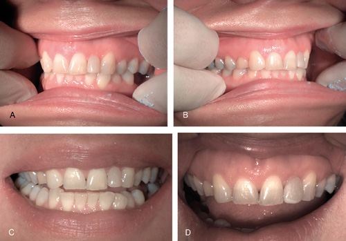
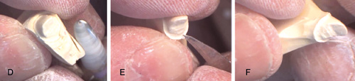
Figur 1. A, B, C, D. Photographs taken with loupe mounted HD-camera sent to the dental technician. Patient was dissatisfied on the asymmetric appearance in the front area as well as on the suboptimal inclination and on the dark color of the second left incisor (A, B, C). According to the wax up the mesial corner of the right canine was restored with composite and a mock up was made on the second left lateral incisor (D).
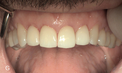
The treatment plan was based on the wax up and the dentist and the patient decided to restore the second left incisor with porcelain crown and fill the mesial corner of the right canine with composite to achieve more symmetric appearance in the front area (fig.1 d, e, f).
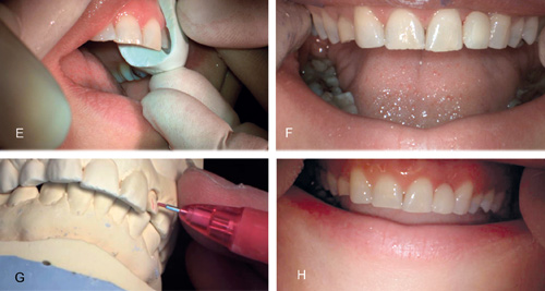
Figur 1. E, F, G, H. Photographs (E, H) and still images of video (F, G) taken with loupe mounted HD-camera. After the preparation of the left lateral incisor the impression was taken and a temporary crown was made (E, F). After the first impression it appeared that the gingival margin was left too coronally and the tooth was not prepared enough on the buccal side. Followed the instructions received in form of a video from the dental technician (G) the treatment continued as planned and the patient was satisfied with the end result (H).
After the first impression it appeared that the gingival margin was left too coronally to achieve more symmetric appearance also in the gingival area. In addition the tooth was not prepared enough on the buccal side (fig.1 g).
All the steps were recorded and videos and photographs were shared between the dentist and the dental technician. The dental technician sent a video with a verbal explanation about areas around the tooth to be prepared to achieve a better esthetic result. Followed the instructions received in form of a video the treatment continued as planned and the patient was satisfied with the end result (fig.1 h).
2. Video used to give feedback to the dental technician.
A dentist started to work with a new dental technician and the first implant-retained crown did not fulfill the expectations of the dentist. The feedback was given with a video combined with a verbal explanation (fig.2). The dental technician remade the implant-retained crown according to the feedback and the teamwork between the dentist and dental technician reached to a higher level.
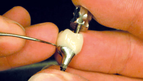
Figur 2. Still image of a video combined with a verbal explanation taken with loupe mounted HD-camera. The implant-retained crown did not fulfill the expectations of the dentist and the feedback was given to the dental technician with a video combined with a verbal explanation.
3. Video used to receive crucial information from the dental technician.
In a prosthetic treatment in the front area the dentist noticed a defect in the impression on the palatal side of the second left lateral incisor. Unfortunately there was no time to remake the impression and the treatment continued as planned (fig 3 a, b, c).
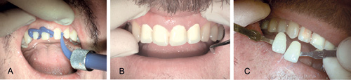
Figur 3. A, B, C. Photographs taken during the operation sent to the dental technician. After the preparation in the frontal area the impression was taken and the temporary crowns was made (A, B). Photographs used to define the color and translucency in the frontal area (C).
After creating a model of a patient's mouth by pouring plaster into the impression the technician examined the model. The dental technician shared the video where he prepared the defect on the plaster cast and managed to find the preparation line reliably (fig 3 d, e, f). The crown was made as planned and the dentist was confident with the fit on the patients tooth (fig 3 g).
Figur 3. D, E, F. Still images of the video the dental technician sent to the dentist taken with loupe mounted HD-camera. The dentist noticed a defect in the impression but there was no time to remake the impression. The dental technician shared the video where he prepared the defect on the plaster cast and managed to find the preparation line reliably (D, E, F).
Figur 3. G. Photograph from the end result sent to the dental technician as a feedback of an excellent work.
Advantages
High quality video material is superior way to communicate:
1. The dental technician has the possibility to see the whole operation. This enables the technician to get more information about the intraoral situation and about the prepared teeth.
2. Still images and videos combined with a verbal instruction shared over the Internet reaches both parts on the same time and doesn't require simultaneous time from the dentist and the dental technician. Comments, questions and additional information can easily be added along the whole procedure.
3. To give or receive feedback with objective video with verbal instructions given over the Internet is effective and it enables to reach to a better teamwork.
4. From a video the resilience of oral mucosa can easily be seen. The dental technician has the possibility to see the movements of lower jaw, lips, smile line, tongue, frenulae, floor of the mouth and soft palate for example.
Discussion
To be able to shoot a high quality photography or video material from the oral cavity is challenging for several reasons. To shoot a video or photographs from an operation it traditionally requires an additional person to hold the video camera or the camera is attached to a lever arm or to the lamp. This leads to a video or photographs witch are not shot from the same angle than the operators view which inevitable leads to shadows and barriers front of the video camera especially in operations made in the back area of the mouth. In addition the video requires editing, which is time consuming, and it requires knowledge of the editing programs. Until this day, it was not possible to attach the video camera on the loupes to be able to capture video and take photographs from the operators view without interruption.
Even if the patient is unrecognizable in the video or in photographs and the recorded material is treated anonymously, the legislation of using this material differs in Nordic countries. Commonly the consent for the use of the recorded material is requested from the patient. For a secure handling or sharing videos or photographs over the Internet, it is essential to use SSL encryption.
According to our preliminary results video material combined with photographs enabled to a better teamwork. Given the instructions using simultaneously dynamic images (video) and verbal instructions in addition to static images (photographs) the dental technician receives significantly more information compared to the information received in a traditional way. Furthermore the technician has the possibility to show to the dentist the specific and essential details related to the case.
Despite the fact that the sample size is quite limited, the results are promising and further studies are suggested. Forthcoming technologies used in the video camera system allow dentist-technician team to reach to a significant higher level of cooperation. In the future the video material will take a significant higher part in the communication in a treatment team.
Acknowledgements
The concept of the Futudent HD camera system is based on Peter Rusanen´s idea to enhance the communication in the dentistry and is a founder of this early state start up company. MDT Mikko Kääriäinen and Prof Kirsi Sipilä don't have any conflict of interests.
English summary
Using video material for communication with the dental technician.
The purpose of this first case report was to evaluate video material as a communication tool with the dental technician. A light loupe mounted HD camera was used to capture the sight of the dentist and the dental technician during the daily work. The verbal instruction was recorded as well. In three representative cases the video material with the verbal instructions and photographs forms an essential part of the communication to enhance the cooperation of the dental technician and the dentist.
Hovedpunkter | |
|---|---|
· |
To minimize the potential for remakes and miscommunication there is a need for a more efficient way to communicate with the dental technician. |
· |
In general video material is rarely used as a daily communication tool. |
· |
From a video the dental technician receives significantly more information of patient's intraoral condition: movements of lower jaw, resilience of the oral mucosa, smile line and color of teeth can easily be seen. |
· |
Video material enhances the communication with the dental technician and a higher level of co-operation is reached. |
Reference
Davenport JC, Basker RM, Heath JR, Ralph JP, Glantz PO, Hammond P. Communication between the dentist and the dental technician. Br Dent J. 2000 Nov 11; 189 (9): 471 - 4.
Griffin JD Jr. Excellence in photography: heightening dentist-ceramist communication. Dent Today. 2009 Jul; 28 (7): 124 - 7.
McLaren EA, Schoenbaum T. Compend Contin Educ Dent. Digital photography enhances diagnostics, communication, and documentation. 2011 Nov-Dec; 32 Spec No 4: 36 - 8.
Sousa Dias N, Tsingene F. SAEF - Smile's Aesthetic Evaluation form: a useful tool to improve communications between clinicians and patients during multidisciplinary treatment. Eur J Esthet Dent. 2011 Summer; 6 (2): 160 - 76.
Schoenbaum TR, Chang YY. Dentist-technician collaboration in the digital age: enhancing outcomes through photography, teamwork, and technology. J Calif Dent Assoc. 2011 Aug; 39 (8): 559 - 67.
Correspondence: Peter Rusanen, Krämertintie 5 as 3, 00620 Helsinki, Finland. E-mail: peter.rusanen@helsinki.fi
Artikkelen har gjennomgått ekstern faglig vurdering.
Rusanen P, Kääriäinen M, Sipilä K. Using video material for communication with the dental technician. Nor Tannlegeforen Tid. 2014; 124: 36-40.
Artikkelen er fagfellevurdert.
Artikkelen siteres som:
Rusanen P, Kääriäinen M, Sipilä K, Rusanen P, Kääriäinen M, Sipilä K. Using video material for communication with the dental technician. Nor Tannlegeforen Tid. 2014;124:36-40. doi:10.56373/2014-1-12